Product Name – Animal Tissue
Quantity/Pack Size – Single Prepared Slide
Form – Tissue Mounted Slide
Grade – Laboratory Grade
Application – Microscopy, Tissue Morphology, Cellular Observation
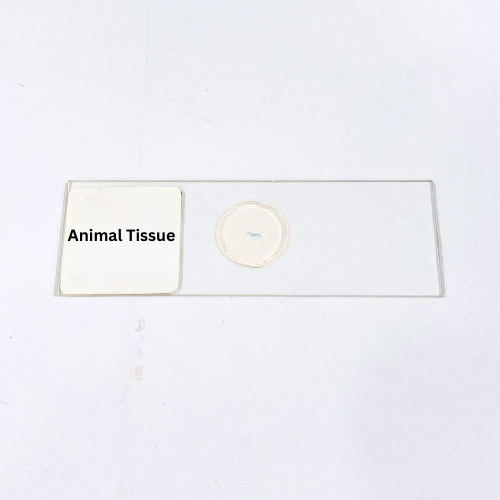
The Animal Tissue slide is prepared with meticulous care to provide a clear, high-resolution view of animal tissue architecture under a microscope. The specimen showcases distinct tissue layers, cellular arrangement, and structural differentiation, highlighting essential components such as connective tissue, epithelial layers, and specialized cellular formations. Precision staining enhances contrast, allowing visualization of intricate details like nuclei, cytoplasm, and extracellular matrix structures. The tissue is securely mounted on a durable glass slide, ensuring long-lasting quality and consistent optical clarity. Its uniform thickness enables accurate focusing and superior light transmission, supporting high-magnification observations. This slide is optimized for detailed study of tissue organization, cellular interrelationships, and structural variations. Prepared under strict laboratory standards, it maintains the integrity of tissue architecture and staining, providing reproducible, reliable results. The Animal Tissue slide serves as an essential tool for observing microscopic tissue details, enabling precise structural analysis, morphological assessment, and comparison across different tissue types. Its durable mounting ensures longevity and usability for repeated examinations without compromising specimen quality. The slide is suitable for various microscopy techniques, delivering sharp images and consistent visualization for in-depth study of tissue structures and cellular organization in professional laboratory environments.
Brightfield and phase-contrast microscopes provide optimal visualization of tissue structures and cellular organization.
Store in a cool, dry area away from direct sunlight and humidity to preserve staining and tissue integrity.
Yes, the specimen is mounted securely for repeated microscopic use without significant degradation.
Yes, other laboratory-prepared slides featuring different animal tissues or specialized staining techniques can be used for comparison.
Hold the slide by its edges or use gloves to prevent contamination and preserve specimen clarity.
Laboratory-grade Vaucheria Reproductive preserved specimen with intact reproductive organs, filamentous thalli, and clear cell structures for precise microscopic morphology studies.
High-quality Leech T.S., laboratory-grade preserved specimen highlighting digestive, circulatory, and nervous systems for precise microscopic morphological examination.
High-quality Liver Fluke Miracidium W.M., laboratory-grade preservation, maintaining structural integrity for accurate microscopic examination of internal and external features.
High-quality Liver Fluke Cercaria W.M., laboratory-grade preservation maintaining anatomical features, suitable for clear microscopic observation of morphology and structural details.
High-quality Balanoglossus preserved specimen, laboratory-grade preparation with maintained anatomical integrity, ideal for detailed marine invertebrate observation and structural analysis.
Product Name – Animal Tissue
Quantity/Pack Size – Single Prepared Slide
Form – Tissue Mounted Slide
Grade – Laboratory Grade
Application – Microscopy, Tissue Morphology, Cellular Observation
The Animal Tissue slide is prepared with meticulous care to provide a clear, high-resolution view of animal tissue architecture under a microscope. The specimen showcases distinct tissue layers, cellular arrangement, and structural differentiation, highlighting essential components such as connective tissue, epithelial layers, and specialized cellular formations. Precision staining enhances contrast, allowing visualization of intricate details like nuclei, cytoplasm, and extracellular matrix structures. The tissue is securely mounted on a durable glass slide, ensuring long-lasting quality and consistent optical clarity. Its uniform thickness enables accurate focusing and superior light transmission, supporting high-magnification observations. This slide is optimized for detailed study of tissue organization, cellular interrelationships, and structural variations. Prepared under strict laboratory standards, it maintains the integrity of tissue architecture and staining, providing reproducible, reliable results. The Animal Tissue slide serves as an essential tool for observing microscopic tissue details, enabling precise structural analysis, morphological assessment, and comparison across different tissue types. Its durable mounting ensures longevity and usability for repeated examinations without compromising specimen quality. The slide is suitable for various microscopy techniques, delivering sharp images and consistent visualization for in-depth study of tissue structures and cellular organization in professional laboratory environments.
Brightfield and phase-contrast microscopes provide optimal visualization of tissue structures and cellular organization.
Store in a cool, dry area away from direct sunlight and humidity to preserve staining and tissue integrity.
Yes, the specimen is mounted securely for repeated microscopic use without significant degradation.
Yes, other laboratory-prepared slides featuring different animal tissues or specialized staining techniques can be used for comparison.
Hold the slide by its edges or use gloves to prevent contamination and preserve specimen clarity.
Laboratory-grade Volvox Daughter Colony W.M. preserved specimen showing intact daughter colonies, spherical colonial arrangement, and individual cell visibility for precise microscopic studies.
High-quality Leech T.S., laboratory-grade preserved specimen highlighting digestive, circulatory, and nervous systems for precise microscopic morphological examination.
High-quality Tapeworm Proglottid Gravid, laboratory-grade whole mount, preserving reproductive organs and segmental structures for precise microscopic observation.
High-quality Heteroneries Parapodia W.M., laboratory-grade whole mount, enabling clear visualization of segmental parapodia and internal structures under microscope.
High-quality Pediculus Head Louse W.M., laboratory-grade preservation maintaining anatomical structure, ideal for detailed microscopic and morphological study.
Agra | Firozabad | Hathras | Mathura | Vrindavan | Aligarh | Lucknow | Ambala | Mainpuri | Shikohabad | Alwar | Bhind | Bharatpur | Morena | Gwalior | Orai | Kanpur | Jhansi | Hardoi | Shahjahanpur | Lakhimpur | Pilibhit | Haldwani | Basti | Gorakhpur | Deoria | Azamgarh | Prayagraj | Varanasi | Sultanpur | Moradabad and more….
Not a member? Create an account
Already got an account? Sign in here
By Clicking “Download Now”, you are confirming that you have read and agree to eqipped.com ‘s Terms of Use and Privacy Policy.
By Clicking “Join Now”, you are confirming that you have read and agree to eqipped.com ‘s Terms of Use and Privacy Policy.