Description
Specifications
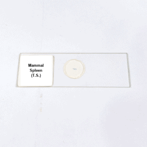
Product Name – Mammal Spleen (T.S.)
Quantity/Pack Size – Standard Pack
Form – Transverse Section Slide
Grade – Laboratory Grade
Application – Microscopic Observation of Splenic Tissue Architecture
Product Overview
The Mammal Spleen (T.S.) slide is meticulously prepared to provide a comprehensive view of splenic tissue in transverse section, highlighting both red and white pulp regions. The slide reveals detailed structural organization, including splenic cords, sinusoids, and lymphoid follicles, enabling clear differentiation of functional zones. High-quality mounting preserves the natural arrangement of cells and vascular channels, ensuring stable and clear observation under a microscope. The white pulp areas are distinctly visible, showing lymphocyte-rich zones, while red pulp demonstrates the filtration network with vascular channels and splenic cords. This precision slide allows examination of the interaction between vascular and lymphoid components, providing insight into the structural complexity of the spleen. The laboratory-grade preparation enhances contrast, allowing for the identification of fine cellular details and tissue boundaries. Durable glass mounting prevents specimen damage and ensures long-term usability for repeated observations. The slide is ideal for those requiring high clarity and consistent structural representation in splenic tissue studies. It offers a reliable tool for detailed microscopic exploration of mammalian spleen architecture, including both functional zones, vascular structures, and cellular composition. With precise preservation and high-quality preparation, this Mammal Spleen (T.S.) slide meets the needs of laboratories seeking dependable and accurate histological specimens for analysis.
FAQs
1. Is this Mammal Spleen slide compatible with standard laboratory microscopes?
Yes, it is compatible with all standard light and compound microscopes for detailed observation.
2. How should the slide be stored to maintain quality?
Store in a cool, dry place away from direct sunlight and moisture to preserve the tissue structure.
3. Does the slide clearly differentiate red and white pulp regions?
Yes, it shows distinct white pulp follicles and red pulp vascular networks with clarity.
4. Can this slide be used multiple times without degradation?
Yes, the durable mounting allows repeated observations without compromising the specimen quality.
5. Are there alternative organ slides available for comparison studies?
Yes, slides of liver, kidney, and thymus are available for comparative histological analysis.


