Description
Specifications
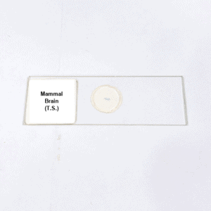
Product Name – Mammal Brain (T.S.)
Quantity/Pack Size – Standard Pack
Form – Slide Specimen
Grade – Laboratory Grade
Application – Microscopic Observation of Brain Tissue
Product Overview
The Mammal Brain (T.S.) slide is prepared with high precision to reveal the structural organization of brain tissue under microscopic observation. The specimen displays clear differentiation between grey matter and white matter regions, with staining methods applied to enhance the visibility of cellular structures. Neurons, glial cells, and vascular arrangements can be observed with clarity, providing a detailed representation of tissue architecture. The transverse section highlights the compact cellular organization and distinct boundaries within brain regions, allowing accurate identification of key tissue features. The preparation process ensures that the tissue morphology is well-preserved, maintaining the integrity of delicate cellular structures. The slide is mounted on durable glass with a stable medium, preventing fading or distortion over time, which supports repeated use. Enhanced staining provides excellent contrast, ensuring that fine structures such as nuclei, cell bodies, and fibers are distinctly visible. The laboratory-grade quality guarantees high-resolution observation across multiple magnifications, maintaining specimen reliability for extended periods. Designed with durability and accuracy in mind, the Mammal Brain (T.S.) slide delivers consistent performance and provides a reliable view of mammalian brain tissue structure for thorough microscopic examination.
FAQs
1. Does the Mammal Brain (T.S.) slide show grey and white matter?
Yes, the slide displays clear differentiation between grey matter and white matter regions of the brain.
2. Is this slide suitable for use with compound light microscopes?
Yes, it is fully compatible with compound light microscopes for detailed tissue observation.
3. How is the specimen preserved on this brain tissue slide?
The specimen is permanently mounted with a protective medium to ensure durability and clarity.
4. What is the recommended way to store this laboratory slide?
It should be stored in a dry, cool place away from sunlight and moisture for long-lasting quality.
5. Are there related nervous system slides available for comparison?
Yes, additional slides such as spinal cord and nerve tissue are available for comparative study.


