Description
Specifications Table
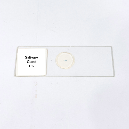
Product Name – Salivary Gland T.S.
Quantity/Pack Size – Single Slide
Form – Microscopic Tissue Slide
Grade – Laboratory Grade
Application – Microscopic Observation of Salivary Gland Tissue
Product Overview
The Salivary Gland T.S. prepared slide is meticulously crafted to provide a high-quality visual representation of salivary gland tissue under a microscope. Each specimen is carefully fixed and mounted using laboratory-grade materials to preserve tissue integrity and ensure clear, detailed observation. The slide is prepared with precise sectioning techniques that maintain the natural structure of acini and ducts, allowing for accurate visualization of cellular arrangements. Protective sealing prevents environmental damage such as moisture, dust, or accidental scratches, ensuring long-term usability. This slide is compatible with a range of magnifications, from low to high power, enabling comprehensive examination of tissue morphology. Its uniform preparation guarantees consistency and clarity across multiple observations, making it a reliable tool for professional laboratory settings. The mounting medium used ensures specimen stability, supporting repeated studies without compromising quality. The Salivary Gland T.S. slide serves as an essential reference for understanding tissue structure and morphology. Its durable construction, professional-grade quality, and precise preparation provide clear visualization, supporting accurate microscopic assessment. Designed for students, researchers, and educators, this slide offers consistent performance, allowing for detailed examination of glandular structures and contributing to a thorough understanding of salivary gland histology.
1. What type of mounting medium is used for the Salivary Gland T.S. slide?
The slide is mounted using a laboratory-grade medium that preserves tissue structure and prevents specimen degradation.
2. Can this slide be used with oil immersion objectives?
Yes, it is compatible with low, high, and oil immersion objectives for detailed microscopic analysis.
3. Are there alternative tissue slides available for comparison?
Yes, other glandular tissue slides such as Pancreas T.S. and Liver T.S. can be used for comparative studies.
4. How should the Salivary Gland T.S. slide be stored?
Store in a protective slide box away from direct sunlight, moisture, and dust to maintain clarity and durability.
5. Is the slide suitable for repeated microscopic observations?
Yes, the professional mounting and sealing allow for multiple examinations without affecting the specimen quality.