Description
Specifications Table
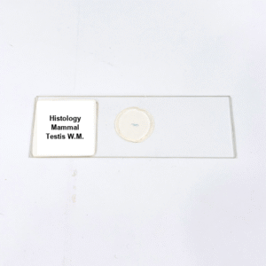
Product Name – Histology Mammal Testis W.M.
Quantity/Pack Size – Single Slide
Form – Mounted Slide
Grade – Laboratory Grade
Application – Observation of Mammal Testis Tissue Structure and Cellular Components under Microscope
Product Overview
The Histology Mammal Testis W.M. slide presents a meticulously prepared, high-quality specimen highlighting the intricate structure of mammalian testis tissue. The slide displays seminiferous tubules in longitudinal and cross-sectional orientations, allowing clear observation of germinal epithelium and associated supporting cells. Spermatogenic cells at various stages, along with Sertoli and Leydig cells, are distinctly visible, demonstrating the cellular organization and functional architecture of testicular tissue. Prepared with laboratory-grade mounting medium, the specimen preserves cell morphology and tubule integrity, ensuring long-lasting clarity and stability. The transparency and uniform thickness of the slide enhance light transmission, facilitating detailed visualization under standard compound microscopes. The specimen is carefully fixed and mounted to prevent distortion or overlapping of cellular structures, providing an accurate representation of testis tissue. The slide highlights the spatial arrangement and density of seminiferous tubules and interstitial tissue, ensuring precise observation of reproductive tissue morphology. Its preservation quality ensures repeated use without degradation of cellular details. This slide is ideal for laboratory studies, enabling precise examination of cellular patterns, tissue architecture, and microscopic features of mammalian testis, supporting reliable observation, documentation, and analysis in research settings. High-quality preparation and consistent clarity make it a dependable resource for microscopic evaluation of testicular tissue organization and cellular composition.
1. How should this Mammal Testis W.M. slide be handled?
Handle the slide by its edges to prevent fingerprints, smudges, or damage to the specimen area.
2. Is the slide compatible with standard laboratory microscopes?
Yes, it is suitable for use with compound, bright-field, and standard laboratory microscopes for detailed tissue observation.
3. Are there alternative slides for reproductive tissue studies?
Alternatives include Histology Mammal Ovary W.M. and Seminiferous Tubule Cross-section slides for comparative reproductive tissue analysis.
4. What is the recommended storage condition for this slide?
Store in a dry, dust-free environment away from direct sunlight and excessive humidity to maintain specimen quality.
5. Where is the Histology Mammal Testis W.M. slide sourced from?
The slide is prepared by certified laboratory suppliers using high-quality materials to ensure accurate preservation and clear visualization of tissue structures.