Description
Specifications Table
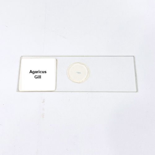
Product Name – Agaricus Gill
Quantity/Pack Size – Single Slide
Form – Prepared Microscope Slide
Grade – Laboratory Grade
Application – Fungal Structure Microscopic Analysis
Product Overview
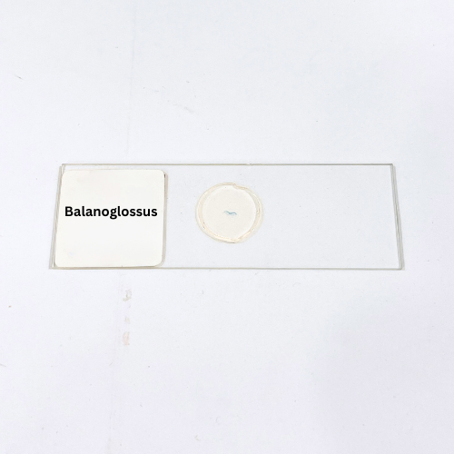
The Agaricus Gill prepared slide features a detailed transverse section of the gill structure from the Agaricus mushroom. It displays lamellae with densely arranged basidia and clearly visible spore-bearing structures, providing an accurate representation of fungal morphology. The slide is mounted under a durable cover glass with a high-quality mounting medium to prevent air bubbles and maintain long-term specimen preservation. Laboratory-grade preparation ensures excellent optical clarity, allowing users to observe fine cellular and structural details, including the arrangement of hyphae and basidiospores. The slide highlights the intricate organization of the gill tissues, enabling precise visualization of spore development patterns and lamellar differentiation. This preparation is ideal for microscopy-focused studies, supporting careful examination of fungal anatomy and cellular structures. The slide is designed for repeated use and can withstand long-term storage without compromising tissue integrity. Its high-quality preparation guarantees consistent results for fungal tissue observation and ensures reliable visual accuracy for laboratory analysis. The Agaricus Gill slide offers a comprehensive and detailed view of mushroom gill morphology, making it a valuable addition for laboratory collections requiring durable, high-clarity specimens for microscopic studies.
1. What magnification is recommended for viewing this slide?
Optimal observation is achieved at 100x to 400x magnification for clear visualization of gill lamellae and basidia.
2. How should the slide be stored to maintain quality?
Store in a dry, dust-free environment away from direct sunlight and extreme temperatures to preserve tissue structure.
3. Is this slide compatible with digital microscopes?
Yes, the slide works well with digital imaging systems for capturing high-resolution images of fungal gill structures.
4. Are there alternative fungal slides available?
Yes, slides for fungal species like Agaricus Cap, Rhizopus, and Albugo are also available for comparative studies.
5. How is the specimen prepared and sourced?
The Agaricus gill tissue is carefully collected and mounted using laboratory-grade methods to ensure accurate structural preservation and clarity.