Description
Specifications
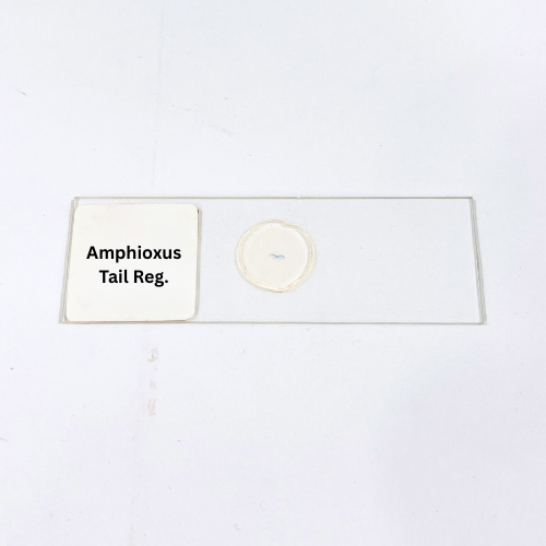
Product Name – Amphioxus Tail Reg.
Quantity/Pack Size – Single Prepared Slide
Form – Mounted Tail Region Section
Grade – Laboratory Grade
Application – Chordate Tail Structure Observation, Microscopy, Tissue Analysis
product overview
The Amphioxus Tail Region slide is a meticulously prepared laboratory-grade specimen that highlights the structural anatomy of the lancelet’s tail. The slide preserves the arrangement of musculature, notochord, nerve cord, and associated connective tissues, providing a comprehensive view of the tail region in a single section. Mounted under a high-quality cover slip, the specimen maintains tissue integrity and staining contrast for long-lasting usability. Special staining differentiates muscular fibers, nerve structures, and connective tissues, allowing detailed observation of cellular and tissue organization. The section is uniform in thickness, ensuring consistent clarity and focus across the entire slide, enabling detailed high-magnification examination. Designed for immediate use, the slide is compatible with various compound microscopes, providing reproducible visualization of the tail’s structural components. Delicate anatomical features, such as segmentation patterns, muscular arrangement, and neural alignment, are preserved for accurate study. The preparation offers durability, clarity, and precise representation, allowing effective observation and exploration of Amphioxus tail structures. With its careful mounting, enhanced staining, and laboratory-grade quality, this slide provides an exceptional tool for microscopic investigation, structural analysis, and comprehensive visualization of the lancelet’s tail anatomy.
FAQs
1. What is the recommended microscope magnification for this slide?
Use high-power objectives between 400x and 1000x to clearly observe muscular and neural structures in the tail region.
2. How should I store this Amphioxus Tail Region slide?
Store in a dry, cool, and dust-free environment to maintain tissue quality and prevent fading of staining.
3. Are there complementary Amphioxus slides available for comparison?
Yes, slides like Amphioxus Intestine, Oral, and Gonad Region are available for comparative tissue analysis.
4. Can this slide be used immediately after purchase?
Yes, it is fully mounted and ready for direct observation without additional preparation.
5. How can I clean the slide without damaging it?
Gently wipe the cover slip with lens paper, avoiding the tissue section to prevent damage or staining loss.