Product Name – Anatropous Ovule
Quantity/Pack Size – Single Slide
Form – Prepared Microscope Slide
Grade – Laboratory Grade
Application – Microscopic Examination of Anatropous Ovule Structure
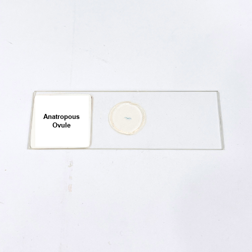
The Anatropous Ovule slide is a meticulously prepared laboratory-grade specimen designed for detailed microscopic observation of ovule structure and orientation. Each slide presents a clear cross-sectional view of the anatropous ovule, showing the nucellus, integuments, and embryo sac arrangement. Precision sectioning ensures thin, uniform slices that facilitate accurate visualization of delicate tissues and internal organization. Advanced staining techniques enhance contrast, highlighting cellular boundaries, nucellar cells, and reproductive structures under various magnifications. Mounted on durable glass, the slide is resistant to mechanical damage, moisture, and common laboratory hazards, ensuring long-term usability. Preparation supports reproducible imaging, providing consistent and reliable observation of ovule morphology. The slide is fully compatible with standard light microscopes, maintaining optical clarity without distortion. Each specimen undergoes rigorous quality control to ensure uniform thickness, precise staining, and faithful representation of anatomical features. The Anatropous Ovule slide balances clarity, durability, and ease of handling, making it a dependable tool for repeated microscopic studies. Professional-grade mounting ensures long-term integrity, enabling multiple observations while maintaining structural and optical quality. With precise preparation and high-quality materials, this slide provides detailed visualization of complex ovule anatomy, supporting clear and accurate laboratory investigations into plant reproductive structures.
It is used for detailed microscopic examination of ovule orientation, nucellus, integuments, and embryo sac.
Yes, it works effectively with all common light and compound microscopes used in laboratories.
Yes, slides like Orthotropous Ovule or Campylotropous Ovule can be used for comparative morphology studies.
Store in a dry, dust-free slide box away from sunlight and humidity to preserve specimen quality.
It is sourced from reputable laboratory suppliers specializing in high-quality prepared microscope slides.
Laboratory-grade Vaucheria Reproductive preserved specimen with intact reproductive organs, filamentous thalli, and clear cell structures for precise microscopic morphology studies.
High-quality Tapeworm Scolex, laboratory-grade preserved specimen highlighting attachment structures, suckers, and hooks for precise microscopic morphological observation.
High-quality Tapeworm Gravid T.S., laboratory-grade transverse section, preserving reproductive and internal structures for precise microscopic observation and study.
High-quality Tapeworm Proglottid W.M., laboratory-grade whole mount, preserving segmental and reproductive structures for precise microscopic examination.
High-quality Liver Fluke Cercaria W.M., laboratory-grade preservation maintaining anatomical features, suitable for clear microscopic observation of morphology and structural details.
Product Name – Anatropous Ovule
Quantity/Pack Size – Single Slide
Form – Prepared Microscope Slide
Grade – Laboratory Grade
Application – Microscopic Examination of Anatropous Ovule Structure
The Anatropous Ovule slide is a meticulously prepared laboratory-grade specimen designed for detailed microscopic observation of ovule structure and orientation. Each slide presents a clear cross-sectional view of the anatropous ovule, showing the nucellus, integuments, and embryo sac arrangement. Precision sectioning ensures thin, uniform slices that facilitate accurate visualization of delicate tissues and internal organization. Advanced staining techniques enhance contrast, highlighting cellular boundaries, nucellar cells, and reproductive structures under various magnifications. Mounted on durable glass, the slide is resistant to mechanical damage, moisture, and common laboratory hazards, ensuring long-term usability. Preparation supports reproducible imaging, providing consistent and reliable observation of ovule morphology. The slide is fully compatible with standard light microscopes, maintaining optical clarity without distortion. Each specimen undergoes rigorous quality control to ensure uniform thickness, precise staining, and faithful representation of anatomical features. The Anatropous Ovule slide balances clarity, durability, and ease of handling, making it a dependable tool for repeated microscopic studies. Professional-grade mounting ensures long-term integrity, enabling multiple observations while maintaining structural and optical quality. With precise preparation and high-quality materials, this slide provides detailed visualization of complex ovule anatomy, supporting clear and accurate laboratory investigations into plant reproductive structures.
It is used for detailed microscopic examination of ovule orientation, nucellus, integuments, and embryo sac.
Yes, it works effectively with all common light and compound microscopes used in laboratories.
Yes, slides like Orthotropous Ovule or Campylotropous Ovule can be used for comparative morphology studies.
Store in a dry, dust-free slide box away from sunlight and humidity to preserve specimen quality.
It is sourced from reputable laboratory suppliers specializing in high-quality prepared microscope slides.
Vaucheria Unsexual culture, supplied in laboratory grade live form, ensures purity, structural clarity, and consistency for reliable microscopic research and precise biological observations.
High-quality Tapeworm Proglottid W.M., laboratory-grade whole mount, preserving segmental and reproductive structures for precise microscopic examination.
Premium Liver Fluke Redia W.M., laboratory-grade whole mount, providing clear visualization of internal structures for precise morphological examination under microscope.
High-quality Sycon L.S. prepared slides, laboratory-grade precision with clear longitudinal tissue sections, optimal staining, and consistent clarity for reliable microscopic observation.
Premium Sycon T.S. prepared slides, laboratory-grade quality with clear tissue sectioning, precise staining, and reliable microscopic visualization for scientific studies.
Agra | Firozabad | Hathras | Mathura | Vrindavan | Aligarh | Lucknow | Ambala | Mainpuri | Shikohabad | Alwar | Bhind | Bharatpur | Morena | Gwalior | Orai | Kanpur | Jhansi | Hardoi | Shahjahanpur | Lakhimpur | Pilibhit | Haldwani | Basti | Gorakhpur | Deoria | Azamgarh | Prayagraj | Varanasi | Sultanpur | Moradabad and more….
By Clicking “Download Now”, you are confirming that you have read and agree to eqipped.com ‘s Terms of Use and Privacy Policy.
By Clicking “Join Now”, you are confirming that you have read and agree to eqipped.com ‘s Terms of Use and Privacy Policy.