Description
Specifications Table
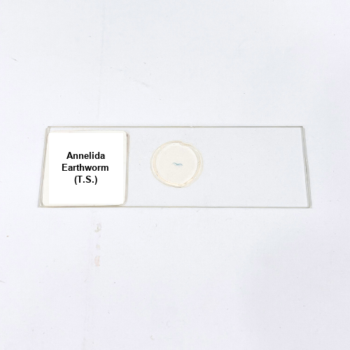
Product Name – Annelida Earthworm (T.S.)
Quantity/Pack Size – Single Slide
Form – Prepared Microscope Slide
Grade – Laboratory Grade
Application – Microscopic Examination of Segmented Worm Tissue Structure
Product Overview
The Annelida Earthworm (T.S.) slide provides a precise transverse section of the segmented worm, enabling detailed observation of internal anatomy. The slide highlights critical features including the epidermis, circular and longitudinal muscle layers, coelomic cavity, nephridia, and alimentary canal. Precision sectioning and staining enhance contrast, making the segmentation, musculature, and tissue layers clearly distinguishable under laboratory microscopes. The slide is mounted on high-quality glass to ensure stability, durability, and clear visualization, allowing repeated use without loss of clarity. Each slide undergoes strict quality control to guarantee uniform staining and structural integrity, providing accurate representation of tissue organization. Protective packaging prevents contamination and physical damage, maintaining long-term usability. The slide allows examination of the coelomic cavity, dorsal and ventral blood vessels, and internal organ arrangement with exceptional detail. It is compatible with standard optical and compound microscopes, ensuring reliable results for observation. The preparation is optimized for clarity, allowing observation of tissue differentiation, segmentation patterns, and cellular arrangement. By maintaining consistent sectioning and staining quality, this slide supports detailed study and reproducibility. Overall, the Annelida Earthworm T.S. slide is an essential tool for observing the complex tissue structure, musculature, and internal organization of segmented worms in a laboratory environment.
1. What structures are visible in the Annelida Earthworm T.S. slide?
The slide shows epidermis, circular and longitudinal muscles, coelomic cavity, nephridia, alimentary canal, and dorsal/ventral blood vessels.
2. Can this slide be used with standard laboratory microscopes?
Yes, it is fully compatible with standard compound and optical microscopes.
3. Are there alternative annelid slides available?
Yes, Lumbricus Cross Section slides and Nereis T.S. slides are available for comparison and study.
4. How should the Annelida Earthworm T.S. slide be stored?
Store in a clean, dry environment, away from direct sunlight and dust to preserve staining and clarity.
5. How is the Annelida Earthworm T.S. slide prepared and sourced?
The slide is carefully sectioned, stained, and mounted using premium laboratory techniques, ensuring durability and reproducible results.