Anthoceros Sporophyte (T.S.)

Anthoceros Sporophyte (T.S.)

Anthoceros Sporophyte (T.S.)

Prepared Anthoceros Sporophyte T.S. slide, laboratory grade, displaying detailed sporophyte structure, cell differentiation, and tissue organization for precise microscopic observation.

Category Brand:

30.00

Specifications Table

Product Name – Anthoceros Sporophyte (T.S.)

Quantity/Pack Size – Single Slide

Form – Prepared Microscope Slide

Grade – Laboratory Grade

Application – Observation of Sporophyte Tissue Structure and Cellular Arrangement

Product Overview

The Anthoceros Sporophyte (T.S.) slide is a high-quality, laboratory-grade preparation meticulously mounted on durable glass for accurate microscopic examination. This transverse section highlights the structural organization of the sporophyte, including the central sporogenous tissue, supportive cells, and surrounding protective layers. Specialized staining techniques enhance the contrast between different cell types, enabling clear identification of spore mother cells, capsule tissues, and surrounding cells. The sectioning process ensures uniform thickness for consistent focus and optimal imaging, while the protective coverslip preserves the specimen from dust, moisture, and mechanical damage. The slide is designed for compatibility with standard compound microscopes, providing sharp resolution and reproducible results. Each Anthoceros Sporophyte slide captures the intricate details of tissue differentiation, cellular arrangement, and reproductive structures, maintaining structural fidelity for extended observation. With careful preparation and staining, the slide offers a clear depiction of sporophyte morphology and internal organization. This ready-to-use slide facilitates detailed study of cell types, tissue boundaries, and overall sporophyte architecture. It is an essential tool for detailed microscopic examination, ensuring clarity, precision, and long-lasting usability for all laboratory observations.

1. What tissues are visible on the Anthoceros Sporophyte slide?

The slide displays sporogenous tissue, capsule cells, and supportive layers clearly for detailed observation.

2. Is this slide compatible with all microscopes?

Yes, it is suitable for standard laboratory compound and optical microscopes.

3. Are there alternative slides for comparison with other bryophytes?

Yes, Marchantia T.S. and Funaria T.S. slides can be used for comparative studies.

4. How should the slide be stored to maintain quality?

Keep in a dry, dust-free environment away from direct sunlight to preserve staining and integrity.

5. How is the Anthoceros Sporophyte slide prepared?

The tissue is carefully sectioned, stained for contrast, mounted, and coverslipped to ensure structural clarity and durability.

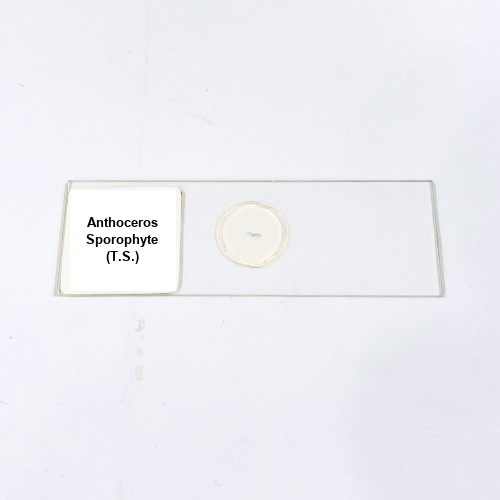
Anthoceros Sporophyte (T.S.)

Anthoceros Sporophyte (T.S.)

30.00

Prepared Anthoceros Sporophyte T.S. slide, laboratory grade, displaying detailed sporophyte structure, cell differentiation, and tissue organization for precise microscopic observation.

Category Brand:

Specifications Table

Product Name – Anthoceros Sporophyte (T.S.)

Quantity/Pack Size – Single Slide

Form – Prepared Microscope Slide

Grade – Laboratory Grade

Application – Observation of Sporophyte Tissue Structure and Cellular Arrangement

Product Overview

The Anthoceros Sporophyte (T.S.) slide is a high-quality, laboratory-grade preparation meticulously mounted on durable glass for accurate microscopic examination. This transverse section highlights the structural organization of the sporophyte, including the central sporogenous tissue, supportive cells, and surrounding protective layers. Specialized staining techniques enhance the contrast between different cell types, enabling clear identification of spore mother cells, capsule tissues, and surrounding cells. The sectioning process ensures uniform thickness for consistent focus and optimal imaging, while the protective coverslip preserves the specimen from dust, moisture, and mechanical damage. The slide is designed for compatibility with standard compound microscopes, providing sharp resolution and reproducible results. Each Anthoceros Sporophyte slide captures the intricate details of tissue differentiation, cellular arrangement, and reproductive structures, maintaining structural fidelity for extended observation. With careful preparation and staining, the slide offers a clear depiction of sporophyte morphology and internal organization. This ready-to-use slide facilitates detailed study of cell types, tissue boundaries, and overall sporophyte architecture. It is an essential tool for detailed microscopic examination, ensuring clarity, precision, and long-lasting usability for all laboratory observations.

1. What tissues are visible on the Anthoceros Sporophyte slide?

The slide displays sporogenous tissue, capsule cells, and supportive layers clearly for detailed observation.

2. Is this slide compatible with all microscopes?

Yes, it is suitable for standard laboratory compound and optical microscopes.

3. Are there alternative slides for comparison with other bryophytes?

Yes, Marchantia T.S. and Funaria T.S. slides can be used for comparative studies.

4. How should the slide be stored to maintain quality?

Keep in a dry, dust-free environment away from direct sunlight to preserve staining and integrity.

5. How is the Anthoceros Sporophyte slide prepared?

The tissue is carefully sectioned, stained for contrast, mounted, and coverslipped to ensure structural clarity and durability.
My Cart
Recently Viewed
Categories

Collaborate With Us

By Clicking “Download Now”, you are confirming that you have read and agree to eqipped.com ‘s Terms of Use and Privacy Policy.

Join Eqipped Circle

By Clicking “Join Now”, you are confirming that you have read and agree to eqipped.com ‘s Terms of Use and Privacy Policy.