Description
Specifications Table
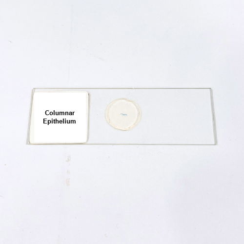
Product Name – Columnar Epithelium
Quantity/Pack Size – Single Slide
Form – Prepared Microscope Slide
Grade – Laboratory Grade
Application – Observation of Cell Structure and Tissue Organization
Product Overview
The Columnar Epithelium slide offers a high-quality, laboratory-grade preparation designed to display the structure and arrangement of column-shaped epithelial cells. The tissue sample is precisely sectioned and stained to enhance contrast, highlighting cell boundaries, nuclei, and cytoplasm for superior visualization. Mounted securely on a durable glass slide with a protective coverslip, the specimen ensures long-lasting use while maintaining the integrity of cellular structures. The slide is prepared to uniform thickness, enabling consistent focus and reducing optical distortions during microscopy. The staining process accentuates cellular features, making it easier to distinguish between individual cells and observe their unique morphology. This slide is ideal for detailed examination of cell alignment, tissue organization, and cellular structure under various magnifications. The mounting medium safeguards the sample from moisture, dust, and environmental factors, ensuring extended usability. Compatible with a broad range of laboratory microscopes, the slide facilitates efficient observation and analysis. Each slide undergoes stringent quality control checks to ensure uniformity and clarity. Whether used for training, research, or diagnostic purposes, this prepared slide meets high laboratory standards, providing precise and accurate visualization of epithelial cell structure for consistent observations and reproducible results in microscopic analysis.
1. What cellular structures can be observed with this slide?
The slide shows cell membranes, nuclei, and cytoplasm with distinct column-shaped arrangements.
2. Can this slide be used with all types of microscopes?
Yes, it is compatible with standard compound and optical microscopes used in laboratory settings.
3. Are there other types of epithelial slides available?
Yes, squamous and cuboidal epithelial slides are available for comparative studies and different observations.
4. How should the slide be stored to ensure longevity?
Store the slide in a dry, dust-free container away from direct sunlight and extreme temperatures.
5. How is the sample prepared for microscopic observation?
The tissue is sectioned thinly, stained for clarity, mounted on glass, and covered with a coverslip for protection.