Description
Specifications Table
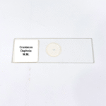
Product Name – Crustacea Daphnia W.M.
Quantity/Pack Size – Single Slide
Form – Whole Mount Slide
Grade – Laboratory Grade
Application – Observation of Crustacean Morphology and Structure
Product Overview
The Crustacea Daphnia W.M. slide is designed for precise and detailed observation of the microscopic structure of freshwater crustaceans. The sample is carefully prepared using whole mount techniques, allowing the intricate anatomy of Daphnia to be viewed clearly under varying magnifications. The slide features excellent light transmission, providing sharp imaging and enhanced contrast that brings out fine details such as appendages, body segments, and internal organ structures. A transparent mounting medium is used to ensure the sample remains stable and protected from external factors such as dust or moisture. The coverslip is tightly sealed to avoid any interference during microscopy sessions, ensuring the longevity of the sample. The specimen preparation follows stringent laboratory standards, ensuring reproducibility and consistency across multiple observations. The slide is compatible with most standard compound microscopes and provides a uniform thickness for accurate focusing. With its robust construction and carefully preserved sample, the Crustacea Daphnia W.M. slide is an indispensable tool for users aiming to explore crustacean morphology. This slide is built to withstand repeated usage while offering clarity and reliability, making it a highly recommended option for anyone requiring detailed structural analysis. Whether for professional or personal research purposes, this product guarantees precise visualization and long-term durability.
1. What details can be observed in this Daphnia slide?
The slide reveals appendages, internal structures, and body segmentation with high clarity under magnification.
2. Is this slide suitable for use with all microscopes?
Yes, it is compatible with standard compound microscopes used for biological specimen observation.
3. Are there similar prepared slides available for study?
Yes, other slides such as Rotifer, Hydra, and Copepod are available for comparative analysis and research.
4. How should this slide be stored to maintain quality?
Store it in a dry, dust-free environment, away from sunlight and high humidity, to prevent damage or fading.
5. How is the sample protected from contaminants?
The slide is sealed with a mounting medium that prevents dust, moisture, and external contaminants from affecting the specimen.