Description
Specifications Table
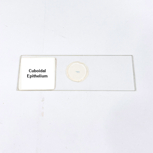
Product Name – Cuboidal Epithelium
Quantity/Pack Size – Single Slide
Form – Mounted Slide
Grade – Laboratory Grade
Application – Observation of Cuboidal Epithelial Cells and Tissue Organization under Microscope
Product Overview
The Cuboidal Epithelium slide offers a high-quality, pre-prepared specimen displaying the typical cuboidal epithelial cells arranged in uniform layers. Each cell maintains its distinct square or cube-like shape with a central nucleus, clearly visible under compound and bright-field microscopes. The slide is prepared using laboratory-grade mounting medium to preserve cellular morphology, prevent distortion, and ensure long-term durability. Transparent mounting allows optimal light transmission, enhancing contrast and providing detailed visualization of cell boundaries and cytoplasmic contents. The slide exhibits the structural organization and compact arrangement characteristic of cuboidal epithelium, enabling precise observation of individual cells and intercellular junctions. This meticulous preparation prevents overlapping or damage to cells, maintaining the natural orientation and clarity of the specimen. The Cuboidal Epithelium slide supports repeated microscopic examinations, offering consistent visibility of nuclei, cell shape, and arrangement. The careful preservation ensures the slide remains intact, making it a reliable tool for observing epithelial tissue morphology. This specimen is essential for detailed study of epithelial structures and can serve as a reference for comparing tissue types, highlighting the orderly arrangement, cell uniformity, and typical cuboidal morphology necessary for accurate structural analysis under a microscope.
1. How should the Cuboidal Epithelium slide be handled?
Handle the slide by its edges to avoid contact with the specimen area, preventing smudges or contamination.
2. Can this slide be used with all standard light microscopes?
Yes, it is compatible with compound and bright-field microscopes for clear observation of epithelial cells.
3. Are there alternative slides for epithelial tissue observation?
Yes, alternatives include Squamous Epithelium and Columnar Epithelium slides for comparative studies of epithelial tissues.
4. What is the recommended storage for this slide?
Store in a dry, dust-free environment away from direct sunlight and high humidity to preserve specimen quality.
5. Where is the Cuboidal Epithelium slide sourced from?
It is prepared by certified laboratory suppliers using high-quality materials for accurate preservation and clear visualization of epithelial cells.