Description
Specifications Table
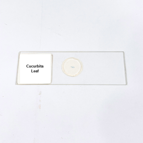
Product Name – Cucurbita Leaf
Quantity/Pack Size – Single Slide
Form – Prepared Microscope Slide
Grade – Laboratory Grade
Application – Leaf Tissue Microscopic Analysis
Product Overview
The Cucurbita Leaf prepared slide offers a detailed transverse and surface view of Cucurbita leaf anatomy, revealing key structures such as the upper and lower epidermis, palisade and spongy mesophyll, stomata, and vascular bundles. Mounted under high-quality cover glass using durable mounting medium, the slide ensures long-lasting preservation of delicate leaf tissues, maintaining clarity without the formation of air bubbles or distortions. Laboratory-grade preparation provides excellent optical clarity, enabling observation of individual cell layers, mesophyll arrangement, and vein patterns. The slide demonstrates differentiation between palisade and spongy parenchyma cells, arrangement of stomata on the epidermis, and clear visibility of xylem and phloem in veins. This ensures precise tissue study and repeated use without degradation over time. The Cucurbita Leaf slide is suitable for a variety of microscopic techniques, including brightfield and digital imaging, and is an essential tool for studying plant leaf morphology and cellular organization. Its meticulous preparation guarantees reproducibility and reliability in observations, offering a consistent visual reference for plant tissue analysis. High-quality mounting and structural integrity make this slide a dependable addition to laboratory collections for detailed microscopic examination of Cucurbita leaf tissue organization.
1. What is the recommended magnification for observing this slide?
Optimal magnification ranges from 100x to 400x for detailed visualization of leaf cells, mesophyll layers, and vascular bundles.
2. How should the Cucurbita Leaf slide be stored?
Keep in a clean, dry environment, away from direct sunlight and humidity to maintain long-term specimen clarity.
3. Can this slide be used with digital microscopes?
Yes, it is fully compatible with both digital and optical microscopes for capturing high-resolution images of leaf tissue.
4. Are alternative plant leaf slides available?
Yes, slides for Dicot Leaf, Monocot Leaf, and other plant species are available for comparative anatomical studies.
5. How is the leaf specimen prepared and sourced?
The Cucurbita leaf is carefully sectioned and mounted using laboratory-grade techniques to ensure clear cellular structure and long-term preservation.