Description
Specifications Table
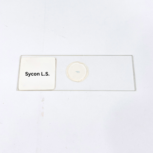
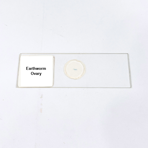
Product Name – Earthworm Ovary
Quantity/Pack Size – 1 Slide
Form – Transverse Section
Grade – Laboratory Grade
Application – Observation of ovary structure and detailed microscopic examination of invertebrate reproductive tissue
Product Overview
The Earthworm Ovary slide presents a meticulously prepared transverse section, highlighting the structure and organization of the ovarian tissue in an earthworm. The specimen is preserved and stained using laboratory-grade techniques, clearly showing ovarian lobes, developing oocytes, and associated connective tissue. The slide ensures exceptional clarity under a microscope, allowing for detailed observation of cell arrangements and tissue morphology. The mounting medium provides long-lasting preservation, maintaining the integrity of delicate reproductive structures while reducing distortion or fading of staining. This slide is ideal for detailed microscopic study of ovary structure, including the distribution of oocytes and supporting tissues. The preparation ensures that even fine cellular features are visible, enabling precise examination and analysis. Each slide is subject to strict quality control, ensuring accurate representation of anatomical features and consistent visibility for laboratory work. The Earthworm Ovary slide offers a reliable, high-quality tool for observing microscopic reproductive structures, supporting in-depth tissue analysis and facilitating a deeper understanding of invertebrate reproductive anatomy in laboratory settings. It is suitable for detailed morphological studies requiring clear, precise visualization of ovary tissues and their organization.
1. What structures can be observed in the Earthworm Ovary slide?
Ovarian lobes, developing oocytes, and connective tissues are clearly visible for detailed microscopic examination.
2. Can this slide be used with standard laboratory microscopes?
Yes, it is fully compatible with conventional optical microscopes for precise tissue observation and analysis.
3. Are there alternative slides for comparative reproductive studies?
Yes, alternatives include Earthworm Testis, Earthworm Seminal Vesicle, and Earthworm Prostate Gland slides for comparative anatomical studies.
4. How should the slide be stored to maintain quality?
Keep in a dry, clean environment away from direct sunlight and moisture to preserve staining and tissue integrity.
5. Where are the specimens sourced from?
Specimens are sourced from certified laboratory suppliers and prepared under strict laboratory-grade protocols for reliability and accuracy.