Description
Specifications
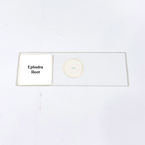
Product Name – Ephedra Root
Quantity/Pack Size – Single Slide
Form – Prepared Microscope Slide
Grade – Lab-Grade
Application – Microscopic Study of Ephedra Root Structure
Product Overview
The Ephedra Root prepared slide is a high-quality specimen designed to provide a clear view of the anatomical features of the root. Each slide is meticulously prepared with precision staining techniques that enhance tissue differentiation, making it possible to observe the organization of the epidermis, cortex, vascular tissue, and medullary regions with exceptional clarity. The slide is mounted on durable glass and sealed securely with a cover slip to prevent contamination and preserve the sample for extended use. Careful attention during preparation ensures consistent staining, uniform section thickness, and minimal interference from air bubbles or mounting defects. This allows for reliable observation at both low and high magnifications under a compound microscope. The Ephedra Root slide highlights important internal structures, including xylem and phloem regions, providing a clear representation of the specimen’s anatomy. The fixation process locks in structural integrity, ensuring that the tissues remain intact over time without distortion. The slide’s stability and precision preparation make it a dependable resource, offering sharp visualization of root structure with long-term durability. Designed with accuracy in mind, it ensures consistent microscopic performance, making it suitable for detailed anatomical studies.
1. What details can be studied from the Ephedra Root slide?
The slide highlights epidermis, cortex, vascular tissue, and medullary regions with clarity.
2. Does the staining provide long-term stability in this slide?
Yes, the slide is stained and sealed to maintain clarity and durability over time.
3. Are other root slides available as alternatives to this?
Yes, prepared slides of roots such as Dicot and Monocot roots are also available.
4. How should the Ephedra Root slide be stored safely?
It should be kept in a dry, cool place away from direct sunlight and moisture.
5. Can this slide be viewed at different magnifications?
Yes, it can be clearly observed under both low and high magnifications using compound microscopes.