Description
Specifications
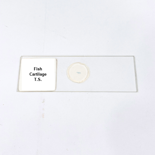
Product Name – Fish Cartilage T.S.
Quantity/Pack Size – Single Slide
Form – Prepared Slide
Grade – Lab-Grade
Application – Observation of Cartilage Structure and Cellular Organization
Product Overview
The Fish Cartilage T.S. prepared slide is a high-quality laboratory specimen designed to display the transverse section of fish cartilage with exceptional clarity. Mounted on a durable optical glass slide and sealed with a protective cover slip, it provides clear visualization of cartilage matrix, chondrocytes, lacunae, and the surrounding connective tissue. Lab-grade preparation ensures uniform thickness and optimal light transmission, allowing precise examination under various magnifications without distortion. Specialized staining enhances tissue contrast, highlighting cellular details and structural organization, making it easy to differentiate between various components of cartilage. The slide is prepared for long-lasting use, preserving the delicate tissue architecture and ensuring consistent clarity over time. It is suitable for researchers, educators, and laboratory professionals seeking a ready-to-use specimen that demonstrates cartilage histology accurately. This slide facilitates detailed observation of tissue morphology and cellular arrangement, supporting high-quality microscopic study and analysis in a laboratory setting.
1. What cartilage features can be observed on this slide?
The slide shows chondrocytes within lacunae, cartilage matrix, and surrounding connective tissues in clear detail.
2. Can this slide be used with standard microscopes?
Yes, it is compatible with all optical microscopes and suitable for detailed observation at multiple magnifications.
3. Are there alternative fish tissue slides available?
Yes, other prepared slides include Fish Bone T.S., Fish Muscle T.S., and Fish Scale W.M. for comparative studies.
4. How should this slide be stored for longevity?
Store in a dry, dust-free slide box, away from direct sunlight and humidity to maintain tissue integrity.
5. What preserves the cartilage structure over time?
High-quality glass, precise staining, and sealed mounting protect the cartilage matrix and chondrocytes for long-term clarity.