Description
Specifications
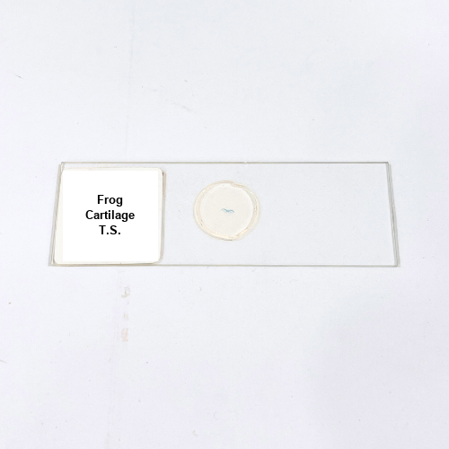
Product Name – Frog Cartilage T.S.
Quantity/Pack Size – Single Slide
Form – Prepared Slide
Grade – Lab-Grade
Application – Observation of Cartilage Tissue Structure and Cells
Product Overview
The Frog Cartilage T.S. prepared slide is a meticulously crafted laboratory specimen designed to provide a detailed view of cartilage tissue structure and cellular composition. The slide is mounted on high-quality optical glass, ensuring clarity and durability for repeated use under various microscope magnifications. The cartilage section reveals chondrocytes within lacunae, embedded in a well-preserved extracellular matrix, highlighting the structural organization of the tissue. Lab-grade preparation guarantees uniform thickness and optimal light transmission, allowing accurate visualization of cellular arrangements and tissue architecture. Specialized staining techniques enhance contrast between cells and matrix, making cellular details easily distinguishable. The slide is carefully sealed to maintain long-term integrity, preventing degradation and ensuring consistent performance over time. Ready-to-use and precise, the Frog Cartilage T.S. prepared slide is an essential tool for laboratory professionals and researchers seeking high-quality visualization of cartilage tissue. It emphasizes cellular and structural details, supporting comprehensive microscopic analysis and facilitating a deeper understanding of connective tissue organization in vertebrates. The preparation ensures reliability, clarity, and durability without the need for additional processing, making it suitable for extensive laboratory observation.
1. What specific tissue structures are visible on this slide?
The slide shows chondrocytes, lacunae, and the extracellular matrix, clearly highlighting cartilage organization.
2. Is this slide compatible with standard microscopes?
Yes, it works with all optical microscopes and supports multiple magnifications for detailed cartilage observation.
3. Are alternative prepared slides available for comparison?
Yes, alternatives include Frog Bone T.S., Frog Heart T.S., and Frog Cartilage W.M. for comparative tissue studies.
4. How should this slide be stored for longevity?
Store in a dry, dust-free slide box away from sunlight and moisture to preserve tissue integrity and clarity.
5. How does the preparation maintain long-term slide quality?
Durable glass, sealed mounting, and specialized staining ensure long-lasting cellular and tissue detail for repeated microscopic use.