Description
Specifications Table
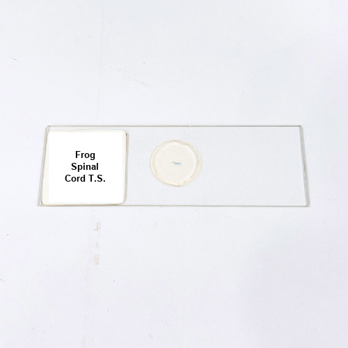
Product Name – Frog Spinal Cord T.S.
Quantity/Pack Size – 1 Slide
Form – Prepared Slide
Grade – Premium Laboratory Grade
Application – Microscopy and neural tissue study
Product Overview
The Frog Spinal Cord T.S. slide is meticulously prepared to provide detailed visualization of amphibian spinal cord anatomy, highlighting neuronal layers, central canal, and surrounding connective tissue. Each sample is preserved with high-quality reagents to maintain cellular integrity and structural accuracy, ensuring long-lasting clarity during microscopic examination. The slide emphasizes critical features such as gray and white matter distribution, nerve fiber arrangements, and supporting tissue morphology, allowing precise study under different magnifications. The mounting medium and cover slip are applied to minimize optical distortion and air bubbles, resulting in superior contrast and sharpness. Premium-grade preparation guarantees consistent results, supporting multiple observations without compromising structural integrity. This slide is fully compatible with standard optical and compound microscopes, making it versatile for various laboratory settings. Careful preservation maintains the slide’s optical properties, ensuring reliable visualization of minute neural details. The Frog Spinal Cord T.S. slide is designed for durability, ease of handling, and extended use, offering a dependable tool for examining amphibian nervous system structures in detail. Special attention to sample handling ensures long-term usability while retaining natural tissue morphology and clarity, providing a high-quality resource for meticulous microscopic observation of spinal cord anatomy.
1. What applications are suitable for Frog Spinal Cord T.S. slides?
They are suitable for microscopic study of amphibian spinal cord structure, including neuronal and connective tissue visualization.
2. Can this slide be used with standard compound microscopes?
Yes, it is compatible with all standard optical and compound microscopes used in laboratory research.
3. Are there alternative slides for comparative neural studies?
Alternatives include Frog Brain T.S. or Frog Nerve Fiber slides for detailed comparative analysis.
4. How should the Frog Spinal Cord T.S. slide be stored?
Store in a cool, dry place away from direct sunlight to preserve tissue structure and optical clarity.
5. Where is this Frog Spinal Cord T.S. slide sourced from?
The slides are obtained from trusted suppliers adhering to strict laboratory quality standards to ensure consistency and reliability.