Description
Specifications
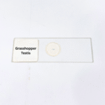
Product Name – Grasshopper Testis
Quantity/Pack Size – Single Prepared Slide
Form – Mounted Tissue Section
Grade – Laboratory Grade
Application – Microscopy, Reproductive Tissue Observation, Cellular Structure Study
product overview
The Grasshopper Testis slide is a premium laboratory-grade specimen designed to provide a comprehensive view of reproductive tissue architecture in insects. The slide features meticulously prepared tissue sections that preserve the organization and morphology of spermatogenic cells, including spermatogonia, spermatocytes, and spermatids, as well as the supporting Sertoli-like structures. The mounting process ensures structural integrity and clarity, allowing for detailed examination under high magnification without distortion. Each slide is carefully stained and mounted to highlight the cellular differentiation and arrangement within the testis, offering consistent and reliable visualization of tissue layers and tubular formations. Ideal for detailed microscopy, the Grasshopper Testis slide enables observation of subtle cellular features and reproductive tissue organization, facilitating precise study of spermatogenic progression. Its laboratory-grade quality ensures long-term stability, preserving morphological details and staining contrast over multiple observations. The slide is ready-to-use, fully mounted, and optimized for ease of handling, making it an essential resource for microscopy, reproductive tissue analysis, and cellular structure study. By providing a clear and accurate representation of insect testis histology, this specimen delivers a high level of precision and reliability for users requiring in-depth examination of reproductive tissues.
FAQs
1. What magnification is best for observing this slide?
High-power objectives ranging from 400x to 1000x are recommended for clear visualization of spermatogenic cells.
2. How should the slide be stored to maintain quality?
Store in a cool, dry place away from direct sunlight and moisture to preserve tissue integrity and staining clarity.
3. Are there alternative slides for reproductive tissue study?
Yes, similar slides include testis sections from other insect species or mammalian reproductive tissues for comparative study.
4. Can this slide be reused multiple times?
Yes, careful handling and proper storage ensure repeated use without compromising tissue structure or staining quality.
5. Is the slide ready for immediate microscopic use?
Yes, it is fully prepared and mounted for immediate observation under a microscope.