Description
Specifications Table
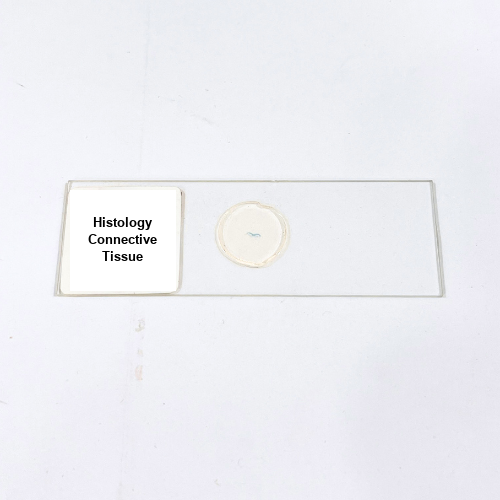
Product Name – Histology Connective Tissue
Quantity/Pack Size – Single Slide
Form – Mounted Slide
Grade – Laboratory Grade
Application – Observation of Connective Tissue Fibers and Cellular Components under Microscope
Product Overview
The Histology Connective Tissue slide provides a meticulously prepared, high-quality specimen exhibiting the intricate organization of connective tissue fibers and cells. The slide demonstrates the arrangement of collagen, elastin, and reticular fibers, along with fibroblasts and other resident cells, clearly visible under compound microscopes. Prepared using laboratory-grade mounting mediums, the specimen maintains cellular integrity and fiber orientation, ensuring long-term durability and minimal distortion. Transparent mounting allows optimal light transmission, providing enhanced contrast and detailed visualization of fibers, cellular components, and extracellular matrix. Each slide is carefully preserved to prevent damage, overlapping, or loss of structural details, making it suitable for repeated microscopic observation. The connective tissue structures are presented in their natural orientation, highlighting the spatial relationship between fibers and cells. The preparation emphasizes uniformity and clarity, allowing accurate observation of connective tissue morphology, fiber density, and cellular distribution. This slide is an essential resource for microscopic studies, enabling precise identification and differentiation of connective tissue components, ensuring consistency in structural visualization. Its preservation quality ensures the specimen remains intact, providing a reliable and detailed view of connective tissue architecture under high magnification, facilitating enhanced observation of fibrous networks and cellular morphology essential for laboratory analysis and microscopic examination.
1. How should the Histology Connective Tissue slide be handled?
Always handle the slide by its edges to prevent fingerprints, smudges, or damage to the specimen area.
2. Is this slide compatible with standard laboratory microscopes?
Yes, it is suitable for use with compound, bright-field, and standard laboratory microscopes for detailed tissue observation.
3. Are there alternative slides for connective tissue study?
Alternatives include Adipose Tissue, Cartilage Tissue, and Bone Tissue slides for comparative histological analysis.
4. What is the recommended storage for this slide?
Store in a dry, dust-free environment, away from direct sunlight and excessive humidity to maintain specimen quality.
5. Where is the Histology Connective Tissue slide sourced from?
The slide is prepared by certified laboratory suppliers using high-quality materials to ensure accurate preservation and clear visualization of connective tissue structures.