Description
Specifications Table
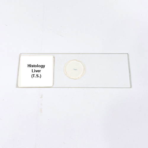
Product Name – Histology Liver (T.S.)
Quantity/Pack Size – 1 Slide
Form – Transverse Section
Grade – Laboratory Grade
Application – Microscopic analysis of liver tissue structure and cellular organization
Product Overview
The Histology Liver (T.S.) slide provides a precisely prepared transverse section of liver tissue, presenting an accurate representation of mammalian hepatic architecture. This slide highlights critical features such as hepatocytes arranged in cords around the central vein, clearly defined sinusoidal spaces, and portal triads, allowing comprehensive observation of tissue organization. The preparation preserves cellular morphology, connective tissue frameworks, and microvascular networks to ensure detailed visualization. The slide employs a superior mounting medium for high contrast and clarity under various magnifications, making it ideal for studying lobular structures, cell distribution, and microanatomy. Its standardized laboratory-grade production ensures durability, reproducibility, and reliable performance for repeated observations. This slide is compatible with conventional optical microscopes and provides consistent, high-resolution imaging of liver tissue features. The attention to detail in preparation allows for clear differentiation of hepatocyte nuclei, cytoplasmic textures, and supporting connective structures. Overall, the Histology Liver (T.S.) slide is a durable, precise, and high-quality tool essential for microscopic tissue analysis, histological research, and morphological studies within laboratory environments.
1. What cellular features can be observed in the Histology Liver T.S. slide?
Hepatocytes, central vein, sinusoidal spaces, and portal triads are clearly visible for detailed tissue analysis.
2. Is this slide compatible with different microscope types?
Yes, it is suitable for standard optical microscopes and ensures high clarity at multiple magnifications.
3. Are there other liver tissue slides available for comparison?
Yes, alternatives include Mammal Liver T.S., fetal liver T.S., and diseased liver T.S. for comparative studies.
4. How should this slide be stored to maintain quality?
Keep in a dust-free, dry environment away from direct sunlight to preserve tissue integrity and clarity.
5. What is the source of the liver tissue used for this slide?
It is sourced from verified laboratory-grade mammalian specimens to ensure accuracy and reliable histological structure.