Description
Specifications
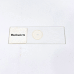
Product Name – Hookworm
Quantity/Pack Size – Single Prepared Slide
Form – Whole Mount Slide
Grade – Laboratory Grade
Application – Parasitic Study, Morphological Observation, Microscopy Analysis
product overview
The Hookworm slide features a meticulously prepared, laboratory-grade parasitic specimen, providing a highly detailed view of its anatomy under a microscope. The slide captures essential morphological structures, including the oral cavity, buccal capsule, esophagus, and attachment structures, allowing for precise examination of its parasitic adaptations. Staining techniques highlight internal and external features, providing contrast for the visualization of reproductive organs, digestive system components, and cuticular patterns. The specimen is mounted securely to preserve structural integrity and prevent degradation over time, ensuring consistent optical clarity and reliable observation. With optimal thickness and transparency, the slide allows efficient light transmission for high-resolution imaging and cellular-level studies. This preparation is designed to offer students, researchers, and educators an accurate representation of the hookworm’s microanatomy. Every detail is preserved, allowing for a comprehensive study of its physical characteristics, including body segmentation and structural adaptations that facilitate parasitic survival. The slide’s durability ensures long-term use in laboratory settings, supporting repeated observation without compromising specimen quality. It serves as a practical and reliable tool for microscopic analysis, offering clear visualization of parasitic features essential for morphological study and detailed research in parasitology.
FAQs
1. What magnification is best for observing the Hookworm slide?
Use 100x to 400x for overall morphology and 400x to 1000x for detailed internal structures.
2. How should the Hookworm slide be stored?
Keep in a cool, dry place away from direct sunlight to maintain staining and tissue quality.
3. Can this slide be compared with other nematode slides?
Yes, comparing with other nematode specimens provides broader understanding of parasitic anatomical variations.
4. Is the Hookworm slide ready to use?
Yes, the specimen is fully mounted and ready for immediate microscopic observation.
5. How should the slide be handled during examination?
Handle by edges with gloves or lens paper to avoid contamination and preserve specimen integrity.