Description
Specifications Table
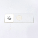
Product Name – Hyaline Cartilage (T.S.)
Quantity/Pack Size – 1 Slide
Form – Transverse Section
Grade – Laboratory Grade
Application – Microscopic study of hyaline cartilage structure and cellular organization
Product Overview
The Hyaline Cartilage (T.S.) slide provides a meticulously prepared transverse section of hyaline cartilage, designed for high-quality microscopic observation. This slide highlights the distinct features of hyaline cartilage, including chondrocytes embedded in lacunae, a smooth extracellular matrix, and uniform fiber distribution. The slide is prepared to preserve tissue integrity, ensuring that cellular morphology and matrix characteristics remain intact for precise study. The mounting medium offers optimal contrast, enhancing the visualization of structural details under various magnifications. This slide is compatible with a wide range of optical microscopes and delivers consistent clarity and image quality. Researchers and educators can observe fine structural arrangements, including lacunar distribution, cell density, and matrix uniformity, supporting detailed morphological assessments. The durability of the slide ensures repeated use without compromising tissue quality. Produced under laboratory-grade standards, the Hyaline Cartilage (T.S.) slide provides reliable performance, clear visualization, and high structural fidelity. It is an essential tool for exploring cartilage architecture, analyzing cellular patterns, and understanding the organization of connective tissue in mammals.
1. What cellular structures are visible on this Hyaline Cartilage T.S. slide?
Chondrocytes in lacunae, extracellular matrix, and fiber distribution are clearly observable under a microscope.
2. Is this slide compatible with different types of laboratory microscopes?
Yes, it works with standard optical microscopes and provides consistent image clarity across magnifications.
3. Are there alternative slides for studying cartilage tissues?
Yes, elastic cartilage T.S. and fibrocartilage T.S. slides are available for comparative histological analysis.
4. How should the slide be stored to maintain quality?
Keep the slide in a dry, dust-free environment, away from direct sunlight to preserve tissue clarity and structure.
5. What is the source of the tissue used in this slide?
The tissue is sourced from verified laboratory-grade specimens to ensure high-quality preparation and accurate morphology.