Description
Specifications Table
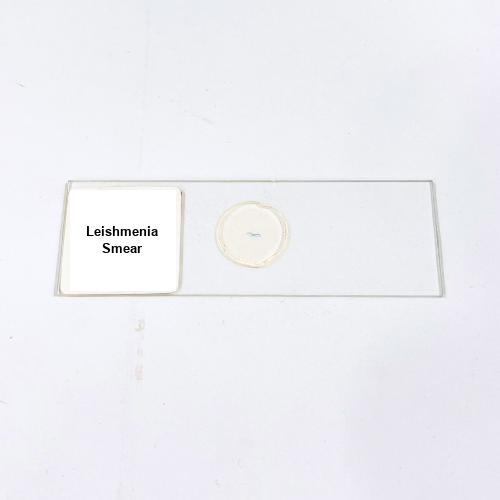
Product Name – Leishmania Smear
Quantity/Pack Size – Single Slide
Form – Prepared Microscopic Slide
Grade – Laboratory Grade
Application – Observation of Leishmania Parasite Morphology and Cellular Structure
Product Overview
The Leishmania Smear slide is a laboratory-grade, meticulously prepared microscopic specimen designed to provide a clear view of the protozoan parasite Leishmania. The slide showcases the distinct cellular morphology, including the nucleus, kinetoplast, and flagellum, allowing for accurate visualization of structural details. The smear is carefully mounted to preserve the integrity of the cells and prevent distortion, ensuring high optical clarity during observation. Prepared with a reliable mounting medium and secured under a durable cover slip, the slide maintains stability for repeated examination under high-power objectives. Its laboratory-grade preparation ensures minimal interference with light transmission, enabling precise study of the protozoan’s cellular arrangement and morphology. This slide allows detailed analysis of parasite shape, internal organelle positioning, and structural variations within different stages of the Leishmania lifecycle. The specimen is suitable for long-term storage, repeated observations, and consistent visualization, providing a dependable resource for laboratory use. The Leishmania Smear slide combines quality preparation, durability, and clarity to facilitate a thorough microscopic examination, highlighting key cellular features and ensuring reliable results for scientific observation and analysis.
1. What is the recommended magnification for viewing this slide?
High-power objectives (40x–100x) are recommended to clearly observe internal organelles and structural details.
2. How should the Leishmania slide be stored?
Keep in a dry, dust-free slide box, away from sunlight and moisture to preserve specimen quality.
3. Are there alternative parasite slides available?
Yes, slides such as Trypanosoma Smear and Plasmodium Smear can be used for comparative protozoan studies.
4. Can this slide be reused for multiple observations?
Yes, the durable mounting medium allows repeated microscopic observations without compromising specimen integrity.
5. What mounting medium is used for this slide?
A laboratory-grade mounting medium is used to maintain cell structure and provide optimal optical clarity.