Description
Specifications Table
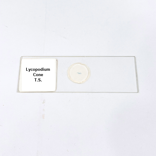
Product Name – Lycopodium Cone T.S.
Quantity/Pack Size – Single Slide
Form – Prepared Microscope Slide
Grade – Laboratory Grade
Application – Microscopic Study of Lycopodium Cone Structure and Reproductive Tissue
Product Overview
The Lycopodium Cone T.S. slide is meticulously prepared to reveal the transverse section of a Lycopodium cone, highlighting its reproductive and structural features. The slide presents clearly organized sporophylls, sporangia, and vascular tissue, providing an in-depth view of the cone’s internal anatomy. Sporangia are distinctly visible, showing their arrangement along the sporophylls, while vascular bundles run through the cone, demonstrating transport pathways and supporting tissue structures. The epidermis and cortex are preserved to show cell morphology and tissue differentiation, allowing observation of cell walls, parenchyma cells, and intercellular spaces. The mounting medium ensures transparency, clarity, and proper preservation of delicate tissues, preventing distortion or overlap. This slide is crafted from high-quality laboratory-grade materials, ensuring durability and consistent visual quality under light and compound microscopes. The Lycopodium Cone T.S. slide facilitates detailed examination of vascular patterns, sporangial arrangement, and overall cone morphology, enabling precise observation of structural complexity. Each slide offers reliable visualization of reproductive tissues and internal anatomy, making it a critical tool for laboratory study of vascular plant cones. The slide provides an accurate representation of the Lycopodium cone’s cellular organization, supporting clear and reproducible microscopic analysis for scientific observation.
1. What structures are visible on the Lycopodium Cone T.S. slide?
The slide shows sporophylls, sporangia, epidermis, cortex, and vascular bundles clearly.
2. Is this slide compatible with standard laboratory microscopes?
Yes, it is compatible with both compound and light microscopes for detailed observation.
3. Are there alternative slides for reproductive tissue study?
Yes, alternatives include Selaginella Cone T.S., Fern Sporangium T.S., and Lycopodium Stem T.S. slides.
4. How should the Lycopodium Cone T.S. slide be stored?
Store in a cool, dry place away from direct sunlight to maintain tissue integrity and clarity.
5. Where is the Lycopodium Cone T.S. slide sourced from?
It is sourced from certified laboratory suppliers ensuring high-quality preparation and consistent reliability.