Description
Specifications Table
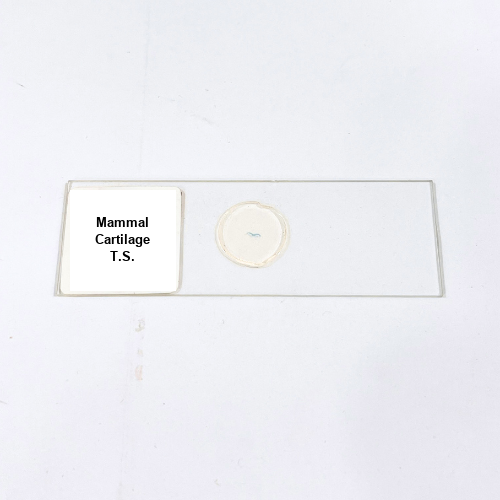
Product Name – Mammal Cartilage T.S.
Quantity/Pack Size – Single Slide
Form – Prepared Slide
Grade – Laboratory Grade
Application – Microscopic Study of Cartilage Connective Tissue Structure
Product Overview
The Mammal Cartilage T.S. prepared slide provides a detailed and high-quality representation of cartilage tissue from mammalian sources, highlighting the arrangement of chondrocytes within the extracellular matrix. Each slide is meticulously fixed and stained to preserve the integrity of the tissue and enhance the contrast between cells and surrounding matrix components. The preparation ensures uniform section thickness, enabling clear visualization of cartilage lacunae, cellular morphology, and matrix distribution under bright-field and phase-contrast microscopy. The slide is mounted with a medium that prevents dehydration, preserves color fidelity, and maintains long-term stability for repeated usage. The careful sealing of the cover slip protects the specimen from mechanical damage, contamination, and deterioration over time. Rigorous quality control is performed to ensure consistency in staining, sectioning, and presentation, providing reliable and reproducible results for laboratory observations. Researchers and laboratory professionals can observe cartilage structure, including cellular and extracellular components, with exceptional clarity. The slide’s optical quality allows for detailed examination of tissue architecture, providing a precise tool for comparative studies and connective tissue research. Its durability and high-quality preparation make it a dependable resource for extended laboratory use, ensuring that the structural features of cartilage are consistently visible and accurately represented.
1. How should the Mammal Cartilage T.S. slide be handled?
Hold the slide by its edges, clean the cover slip if necessary, and place it carefully on the microscope stage for observation.
2. Is this slide compatible with various microscopy techniques?
Yes, it is compatible with bright-field, phase-contrast, and differential interference contrast microscopy for detailed tissue visualization.
3. What are alternative slides for connective tissue studies?
Alternatives include Mammal Bone T.S. and Mammal Tendon T.S. slides for comparative structural analysis of connective tissues.
4. How should the slide be stored after use?
Store the slide in a dry, dust-free environment, away from direct sunlight and humidity to maintain clarity and longevity.
5. Where is this slide sourced from?
The slide is sourced from certified laboratory suppliers and prepared under strict standards to ensure high-quality, consistent specimens.