Description
Specifications Table
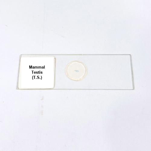
Product Name – Mammal Testis (T.S.)
Quantity/Pack Size – 1 Slide
Form – Transverse Section
Grade – Laboratory Grade
Application – Microscopic study of testis structure, seminiferous tubules, and spermatogenic cell arrangement
Product Overview
The Mammal Testis (T.S.) slide presents a carefully prepared transverse section of mammalian testis tissue, revealing detailed cellular and structural organization. The slide highlights seminiferous tubules, showing the full spectrum of spermatogenic cells, including spermatogonia, spermatocytes, spermatids, and mature spermatozoa. Interstitial cells of Leydig and connective tissue are clearly distinguishable, providing a complete overview of the testis microarchitecture. Prepared using laboratory-grade mounting techniques, the slide ensures optimal preservation of tissue integrity, enhancing visibility under standard optical microscopes. The uniform section thickness allows for accurate observation of tubular arrangement and cellular distribution. The slide also demonstrates the organization of the tubular lumen and surrounding connective tissue matrix, offering a comprehensive view of testicular histology. Durable and standardized, this Mammal Testis (T.S.) slide maintains clarity and structural fidelity over repeated examinations, making it a reliable tool for histological studies. Its high-resolution presentation facilitates detailed tissue analysis, supporting research and laboratory work requiring precise microscopic visualization of testis morphology, cellular composition, and tissue organization, ensuring consistent results in experimental and observational studies.
1. What structures are visible in the Mammal Testis T.S. slide?
Seminiferous tubules, spermatogenic cells, interstitial cells of Leydig, connective tissue, and tubular lumen are clearly visible.
2. Can this slide be viewed using standard microscopes?
Yes, it is compatible with conventional optical microscopes for clear visualization at multiple magnifications.
3. Are there alternative reproductive tissue slides available?
Yes, options include mammal ovary T.S., epididymis T.S., and spermatozoa preparations for comparative studies.
4. How should the Mammal Testis T.S. slide be stored?
Store in a dry, clean environment, protected from direct sunlight and moisture to preserve tissue integrity and clarity.
5. What is the source of the testis tissue used in this slide?
It is sourced from verified mammalian specimens prepared to laboratory-grade standards for accurate histological representation.