Description
Specifications
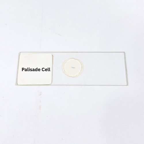
Product Name – Palisade Cell
Quantity/Pack Size – Single Prepared Slide
Form – Plant Tissue Section Slide
Grade – Laboratory Grade
Application – Plant Anatomy, Photosynthesis Study, Microscopy, Cellular Structure Analysis
product overview
The Palisade Cell slide is a high-quality laboratory-grade preparation showcasing the densely packed, elongated photosynthetic cells of plant leaves. Each cell is preserved with exceptional clarity, highlighting prominent chloroplasts and cell walls to facilitate detailed microscopic study. The slide provides a transverse and longitudinal view of palisade tissue, allowing observation of cell arrangement, intercellular spaces, and chloroplast distribution. Optimal staining emphasizes cellular differentiation, making it easier to identify nuclei, chloroplasts, and other subcellular structures. The durable mounting medium ensures long-term preservation, preventing fading or damage to tissue morphology. This slide is suitable for high-resolution imaging, detailed microscopic examination, and comparative analysis of plant leaf structures. Its consistent thickness and transparency enhance optical quality, enabling accurate observation of cell shape, size, and spatial organization. The Palisade Cell slide is an essential specimen for students, educators, and researchers who require precise visualization of plant anatomy, supporting clear understanding of photosynthetic tissue architecture. With meticulous preparation and reliable preservation, this slide provides a dependable resource for laboratory analysis and microscopy exploration.
FAQs
1. Which magnification is suitable for viewing the Palisade Cell slide?
Use 100x to 400x for general tissue arrangement and 400x to 1000x for detailed observation of chloroplasts and cell structures.
2. How should this slide be stored to maintain quality?
Store in a cool, dry place away from direct sunlight to prevent staining deterioration and cellular damage.
3. Can this slide be compared with other plant tissue slides?
Yes, slides like Spongy Mesophyll or Leaf Epidermis provide complementary views for comparative plant anatomy studies.
4. Is the Palisade Cell slide ready for immediate use?
Yes, it is fully mounted and ready for direct microscopic examination without additional preparation.
5. How should the slide be handled during observation?
Handle by edges using lens paper or gloves and avoid touching the specimen to preserve tissue integrity.