Description
Specifications Table
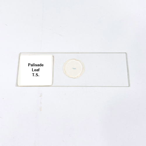
Product Name – Palisade Leaf T.S.
Quantity/Pack Size – Single Slide
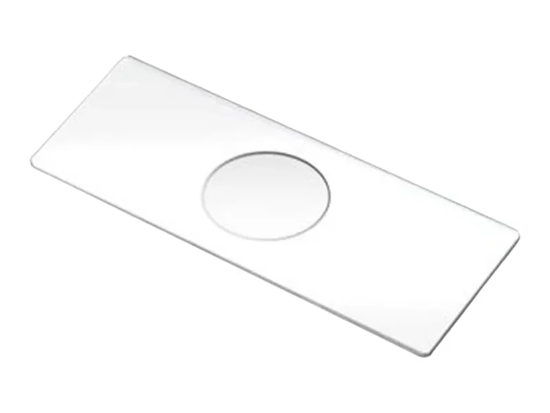
Form – Prepared Microscope Slide
Grade – Laboratory Grade
Application – Leaf Cell Structure Observation
Product Overview
The Palisade Leaf T.S. prepared slide is crafted to offer exceptional clarity and precision for observing leaf tissue structures under the microscope. Utilizing high-grade materials and advanced preparation techniques, the slide presents the palisade cells with minimal distortion and optimal transparency. The leaf tissue is carefully sliced and mounted to preserve cellular integrity, enabling detailed visualization of cell walls, chloroplast distribution, and intercellular spaces. The slide is sealed with a protective cover glass, ensuring long-lasting use and protection against dust, moisture, and external contaminants. Designed for compatibility with standard laboratory microscopes, the slide delivers uniform contrast, making it easier to study the arrangement and morphology of palisade cells. The preparation ensures that cellular components are visible with precision, offering consistent results across multiple observations. The slide’s durability and protective coating enhance usability and make handling and storage convenient. Additionally, the high-quality mounting medium prevents bubbles and refractive distortions, contributing to a seamless viewing experience. Suitable for repeated use, the slide maintains its clarity over time while resisting damage from environmental factors. The Palisade Leaf T.S. slide is a reliable tool for in-depth examination of plant tissues, providing reproducible results and superior optical performance. Its robust construction and meticulous preparation make it an indispensable resource for scientific exploration, allowing for detailed study of leaf anatomy with unparalleled clarity and precision.
1. Can this slide be used with any microscope?
Yes, it is compatible with standard optical microscopes equipped with adjustable magnification settings for observing cellular details.
2. How should the slide be stored to prevent damage?
The slide should be kept in a dry and dust-free container, away from direct sunlight and moisture to preserve its clarity.
3. Are there alternatives for studying leaf structures?
Yes, there are other prepared slides featuring different leaf tissues and cross-sections to explore various aspects of plant anatomy.
4. What is the best way to handle this slide during observation?
Always hold the slide by its edges and avoid touching the mounted tissue area to prevent smudging and contamination.
5. Where is the plant tissue sourced from for this slide?
The tissue is obtained from healthy plant specimens under controlled conditions to ensure authenticity and optimal sample preparation.