Description
Specifications
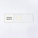
Product Name – Pituitary Gland T.S.
Quantity/Pack Size – Single Prepared Slide
Form – Mounted Tissue Section
Grade – Laboratory Grade
Application – Endocrine Tissue Observation, Microscopy, Cellular Structure Analysis
product overview
The Pituitary Gland T.S. slide is a carefully prepared laboratory-grade specimen designed to showcase the intricate architecture of the mammalian pituitary gland. The tissue is precisely sectioned and mounted to reveal both the adenohypophysis (anterior lobe) and neurohypophysis (posterior lobe) with distinct cellular layers and glandular organization. High-quality staining enhances the contrast between different cell types, allowing clear visualization of chromophobes, acidophils, and basophils in the anterior lobe, as well as nerve fiber terminals in the posterior lobe. The slide’s uniform thickness ensures consistent optical clarity across the entire section, supporting detailed microscopic examination at various magnifications. Observers can analyze glandular lobes, detect cellular morphology, and study the arrangement of supporting connective tissue with precision. The mounting medium preserves tissue integrity and maintains staining stability over time, providing a reliable long-term specimen. Designed for superior clarity and durability, this slide enables meticulous study of pituitary structure, offering a comprehensive view of endocrine organization, cell density variations, and tissue boundaries. With laboratory-grade quality and meticulous preparation, the Pituitary Gland T.S. slide provides an exceptional tool for detailed visualization of complex glandular anatomy and cellular composition, facilitating accurate and repeatable microscopic observations.
FAQs
1. Which microscope magnification is ideal for observing this slide?
High-power objectives between 400x and 1000x are recommended for detailed examination of glandular and cellular structures.
2. How should the slide be stored to preserve quality?
Keep the slide in a cool, dry, and dust-free environment to maintain staining and tissue integrity.
3. Are there alternative slides for comparing glandular tissue?
Yes, other endocrine tissue slides such as adrenal gland or thyroid gland sections can be used for comparative studies.
4. Is the slide ready for immediate use?
Yes, it is fully mounted and can be used directly under a microscope without additional preparation.
5. How can the slide be cleaned safely?
Wipe the edges gently with lens paper; avoid touching the mounted tissue to prevent damage to the specimen.