Description
Specifications Table
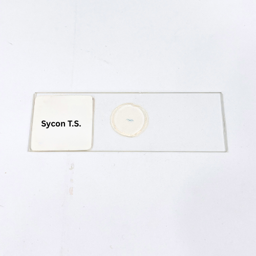
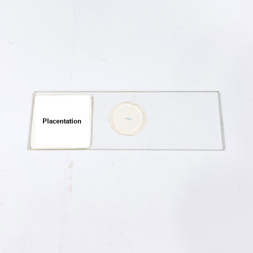
Product Name – Placentation
Quantity/Pack Size – Single Slide
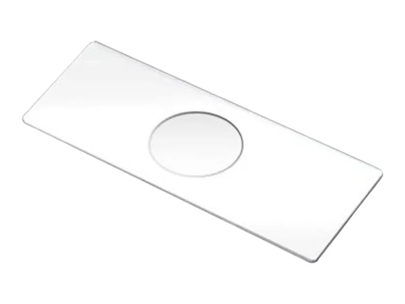
Form – Prepared Slide
Grade – Laboratory Grade
Application – Microscopic Study of Placental Structures
Product Overview
The Placentation prepared slide is meticulously crafted to provide exceptional clarity and structural detail for the examination of placental tissues. The sample is prepared from high-quality biological materials to ensure a representative cross-section that reveals critical features of placental development. Advanced staining techniques are applied to highlight cellular structures, enabling clear differentiation of tissue layers and vascular networks. The slide is mounted using specialized media that maintains sample integrity while preventing drying or contamination, ensuring long-term usability. The coverslip is carefully sealed to reduce the presence of air bubbles and preserve sample clarity, allowing researchers to focus on the microscopic architecture without visual distortions. Compatible with a wide range of microscopes, particularly bright-field setups, this slide supports detailed analysis with high magnification lenses. Its durability ensures that repeated observations do not compromise the staining or structural features. The slide’s uniform thickness minimizes overlapping and provides consistent results across different observations. Additionally, the sample’s staining properties are engineered to remain stable, reducing fading and ensuring clarity over extended periods. Designed to meet rigorous laboratory standards, the Placentation slide offers a reliable and precise medium for biological exploration, supporting clear visualization of complex structures with accuracy and consistency.
1. Can this slide be used with different microscopy techniques?
This slide is compatible with bright-field and phase-contrast microscopy for detailed observation at various magnifications.
2. How should the slide be handled to prevent damage?
Handle the slide by its edges using clean gloves and avoid touching the central specimen area to maintain clarity.
3. Are there other related slides available for comparative studies?
Yes, slides focusing on various developmental stages and tissue types are available for broader biological analysis.
4. What is the recommended storage condition for this slide?
Store it in a dry, dust-free container at room temperature, away from direct sunlight and high humidity to preserve staining quality.
5. Where is this slide sourced from?
It is sourced from certified labs that follow strict preparation and quality control protocols to ensure accuracy and reliability.