Description
Specifications Table
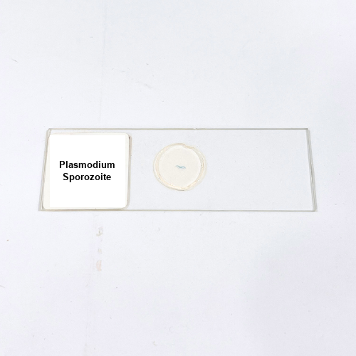
Product Name – Plasmodium Sporozoite
Quantity/Pack Size – Single Slide
Form – Prepared Slide
Grade – Laboratory Grade
Application – Microscopic Observation of Protozoan Structures
Product Overview
The Plasmodium Sporozoite prepared slide is crafted to offer detailed microscopic visualization of this critical stage in the Plasmodium life cycle. Manufactured with precision, the slide presents a high-grade sample that preserves the delicate structures and morphology of the sporozoites. The mounting process is designed to prevent any distortion, ensuring that the cellular and subcellular features remain intact and clear for prolonged observations. The slide is treated to withstand repeated examinations, maintaining its optical clarity without fading or damage. Its compatibility with various microscopy techniques, such as bright-field, phase-contrast, and fluorescence, enables enhanced study of the sporozoite’s form and internal components. The uniformity in preparation ensures reproducibility, allowing users to conduct consistent observations across multiple sessions. The sample is securely sealed to prevent contamination or dehydration, further improving the longevity and usability of the slide. Each slide undergoes strict quality control to meet laboratory standards, offering dependable results for scientific exploration. The optical properties of the slide facilitate high-resolution imaging, making it easier to identify distinct structures within the protozoan. With its robust construction and fine details, this prepared slide stands as an essential tool for microscopic study, providing users with a reliable and clear view of Plasmodium Sporozoite formations under varied laboratory conditions.
1. How should this slide be handled during microscopy?
Handle the slide by its edges using clean gloves to avoid smudges and ensure clear viewing of the specimen.
2. Can this slide be used with advanced imaging systems?
Yes, it is compatible with advanced systems such as fluorescence and phase-contrast microscopes for enhanced imaging.
3. Are there alternative slides for studying protozoans?
Slides like Trypanosoma or Giardia specimens offer additional options for comparative protozoan analysis under microscopes.
4. What storage conditions are recommended for this slide?
Store it in a cool, dry place away from direct sunlight and moisture to maintain clarity and prevent damage.
5. Where is this slide sourced from?
The slide is sourced from certified laboratory suppliers who follow strict preparation and quality assurance processes.