Description
Specifications Table
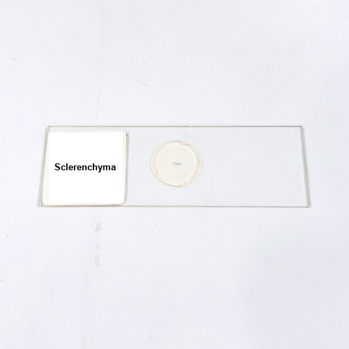
Product Name – Sclerenchyma
Quantity/Pack Size – Single Slide
Form – Prepared Microscope Slide
Grade – Laboratory Grade
Application – Plant Tissue Microscopic Analysis
Product Overview
The Sclerenchyma prepared slide offers a detailed view of plant structural tissues, particularly thick-walled cells, fibers, and lignified components, crucial for microscopic examination. The specimen is mounted carefully under high-quality cover glass with a durable medium, ensuring long-lasting clarity and minimal distortion. Laboratory-grade preparation guarantees optimal optical performance, minimizing air bubbles and providing precise visualization of cell walls, fiber bundles, and associated lignification patterns. This slide highlights the distinct morphology of sclerenchyma cells, including their rigidity, shape, and arrangement within plant tissues. Its meticulous preparation allows reproducible observations, making it suitable for brightfield and digital microscopy. The slide ensures structural integrity, enabling repeated microscopic studies of plant mechanical tissues. With high-quality mounting and consistent specimen presentation, this slide supports accurate analysis of fiber distribution, cell wall thickness, and overall sclerenchyma arrangement. Users can examine characteristic features of sclerenchyma cells, such as elongated fibers and stone cells, with clarity and precision. The slide is durable, suitable for long-term laboratory use, and provides reliable visualization under standard magnifications. By offering a clear and consistent reference, it facilitates detailed exploration of plant structural tissues for research and analytical purposes in laboratory settings.
1. What magnification is recommended for observing Sclerenchyma?
Optimal magnification ranges from 100x to 400x for clear visualization of cell walls and fiber structures.
2. How should this slide be stored to preserve quality?
Keep in a dry, dust-free environment away from direct sunlight and excessive humidity to maintain specimen integrity.
3. Is this slide compatible with digital microscopes?
Yes, it can be used with both optical and digital microscopes for high-resolution imaging of plant tissue structures.
4. Are there alternative slides for comparative plant tissue studies?
Yes, slides such as Parenchyma, Collenchyma, and Xylem tissues are available for comparative microscopic examination.
5. How is the Sclerenchyma specimen prepared and sourced?
The plant tissue is carefully sectioned, fixed, and mounted using laboratory-grade techniques to preserve cell wall integrity and morphology.