Description
Specifications Table
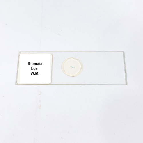
Product Name – Stomata Leaf W.M.
Quantity/Pack Size – Single Slide
Form – Prepared Microscope Slide
Grade – Laboratory Grade
Application – Microscopic Examination of Leaf Stomatal Structures
Product Overview
The Stomata Leaf W.M. slide provides a high-quality, carefully prepared specimen of a leaf epidermis, showcasing the stomatal apparatus in detail. The slide highlights key features including stomatal pores, guard cells, subsidiary cells, and epidermal cell arrangement. Precision mounting and expert staining techniques enhance contrast, allowing clear visualization of the stomatal structure and surrounding cells. The slide is mounted on durable glass to ensure long-term clarity, resistance to damage, and easy handling. Each slide undergoes quality checks to ensure uniform staining, preservation of cellular integrity, and consistent microscopic visibility. Protective packaging maintains slide quality during transport and storage, shielding it from dust, moisture, and contamination. The slide supports detailed observation of the leaf’s epidermal architecture, guard cell morphology, and stomatal distribution, providing reliable and reproducible visualization. Compatible with standard laboratory microscopes, this slide allows accurate examination of cellular anatomy and structural arrangements at high magnification. Its professional preparation ensures precise alignment of the specimen and optimal contrast for microscopic study, making it an essential tool for laboratory microscopy focused on stomatal and epidermal cell features. The slide facilitates detailed, long-lasting observation with superior clarity, reproducibility, and structural accuracy.
1. What features of the leaf can be observed in the Stomata Leaf W.M. slide?
The slide shows stomatal pores, guard cells, subsidiary cells, and epidermal cell patterns.
2. Is this slide compatible with standard microscopes?
Yes, it works with all standard compound and optical laboratory microscopes.
3. Are there alternative prepared leaf slides available?
Yes, slides like Hydrilla Leaf W.M. and Onion Peel W.M. are available for comparison.
4. How should the Stomata Leaf W.M. slide be stored?
Store in a dry, dust-free location, away from direct sunlight to maintain clarity and prevent damage.
5. How is the Stomata Leaf W.M. slide prepared and sourced?
The slide is carefully prepared, stained, and mounted using laboratory-grade techniques ensuring preservation of stomatal and epidermal structure.